Wednesday, August 15, 2012
Fever of Unknown Origin
Today in Morning Report, we discussed the workup of a Fever of Unknown Origin (FUO).
To review, FUO is defined as a temperature higher than 38.3°C on several occasions and lasting longer than 3 weeks, with a diagnosis that remains uncertain after 1 week of investigation, that commonly include:
- a full history and physical
- CBC, electrolytes, creatinine, glucose, liver enzymes, LDH, bilirubin
- blood cultures and blood film
- hepatitis serology (if liver enzymes abnormal)
- urinalysis, urine microscopy, and urine culture
- CXR
The three broad categories in which to frame one's differential diagnosis of FUO commonly include:
- infections
- malignancies
- connective tissue diseases
A systematic review published right here in Toronto in the Archives of Internal Medicine in 2003 detailing a Comprehensive Evidence-Based Approach to Fever of Unknown Origin recommend the following workup for FUO (in addition to the above):
- ANA, RF
- HIV, CMV, EBV, Q-fever
The following diagram outlines a suggested evidence-based approach for the further workup of FUO after the above investigations are complete:
The aim is to find the cause, in order to temper the Disco Inferno.
Friday, August 10, 2012
Viral Meningitis
Today, in Morning Report, we discussed the diagnosis of viral meningitis.
How opportune a moment to firstly review a previous post last month on Bacterial Meningitis.
We discussed the importance of not delaying administration of parenteral medications (antimicrobial therapy +/- dexamethasone) upon suspicion of bacterial meningitis following collection of blood cultures in order to perform a lumbar puncture.
In fact, administration of antibiotics does not change the composition of the CSF cell count, differential, or biochemistry for at least 24 hours. Please click on this link for the abstract to a paper published in 1983 detailing this finding.
However, if one is able to obtain CSF in a safe and timely manner that does not delay patient care, it is certainly preferable, as we recognize from studies, such as this one, that CSF sterilization may occur more rapidly after initiation of parenteral antibiotics than previously suggested, with complete sterilization of meningococcus within 2 hours and the beginning of sterilization of pneumococcus by 4 hours into therapy.
The classic CSF cell count, differential, and biochemistry compositions of different diagnosis can be reviewed here.
Lastly, the most common causes of viral meningitis include enteroviruses, West Nile Virus, and Herpes Simplex Virus (more often type 2).
Thursday, August 2, 2012
Pulmonary Function Tests
Today in Morning Report, we reviewed an approach to pulmonary disease, with a focus on the utility of pulmonary function tests.
And, lastly, here's a link to the ATS/ERS Task Force: Standardization of Lung Function Testing guide for Interpretative strategies for Lung Function Tests published in Eur Respir J in 2005.
I thought this would be an opportune time to review the definitions of the different "capacities" and "volumes" in pulmonary physiology.
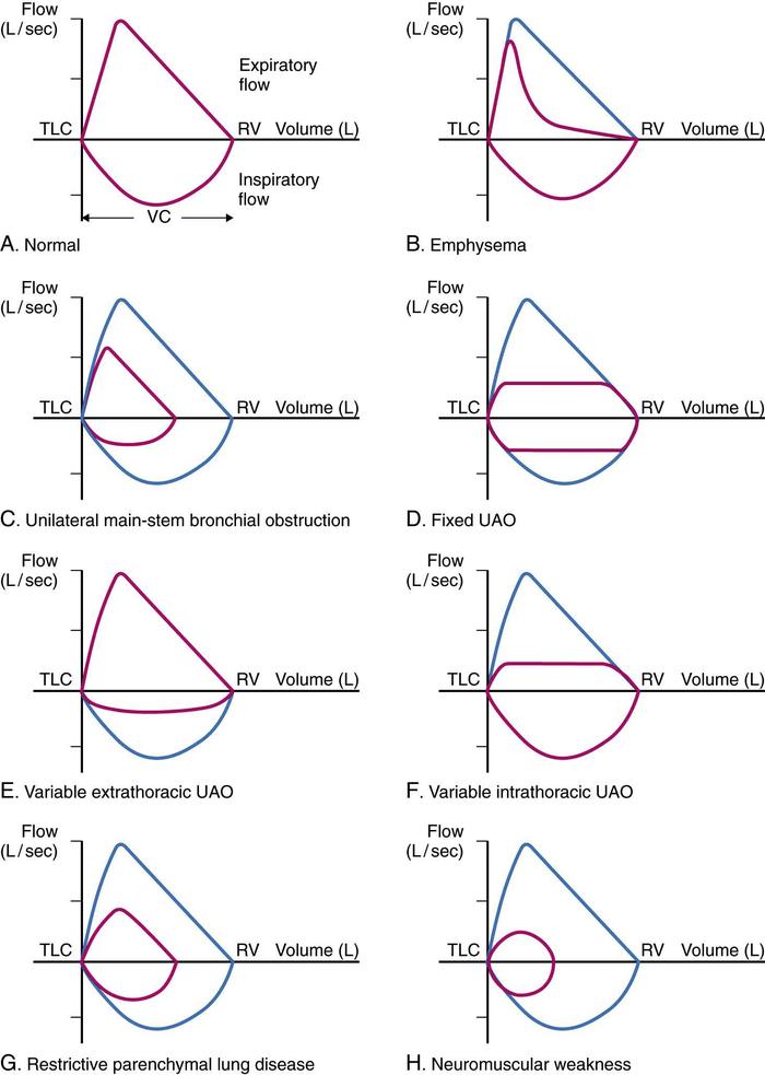
Here is a review of some of the classic flow- volume loop morphologies for pulmonary function tests.
And, lastly, here's a link to the ATS/ERS Task Force: Standardization of Lung Function Testing guide for Interpretative strategies for Lung Function Tests published in Eur Respir J in 2005.
Wednesday, August 1, 2012
Posterior Reversible Encephalopathy Syndrome
Today in Morning Report, we talked about Posterior Reversible Encephalopathy Syndrome (PRES), which is also known under the moniker, Reversible Posterior Leukoencephalopathy Syndrome (RPLS).
PRES is a neurologic syndrome diagnosed by clinical AND radiologic findings:
Clinical Presentation
Classically characterized by the following:
- headaches
- altered level of consciousness
- visual changes
- seizures
Radiologic Findings
The classic findings upon neuroimaging of PRES is symmetrical white matter edema in the posterior cerebral hemispheres, particularly the parieto-occipital regions, although this not always the case. PRES can often been seen on CT imaging of the head, although MRI brain is best.
Here's a CT head non-contrast revealing bilateral hypodensities involving the occipo-parietal region.
Here's a T2-weighted MRI brain demonstrating multiple cortico-subcortical areas hyperintense lesions involving the occipital and parietal lobes, as well as the pons.
Causes of PRES
The most common causes of PRES include:
- hypertensive encephalopathy
- eclampsia
- immunosupprseive medications, including calcineurin inhibitors (cyclosporine, tacrolimus)
- TTP/HUS
- vasculitis (e.g. SLE, PAN)
- renal failure (acute and/or chronic)
Management of PRES
- The most important aspect of management is to treat the underlying cause (whether this means blood pressure lowering, delivery of baby, discontinuation of offending medication, plasmapheresis, etc.)!
- Seizures can be treated with antiepileptic agents, most commonly phenytoin.
Most patients demonstrate the "R" of PRES, meaning their symptoms are reversed by treating the underlying cause, often with two weeks of management, although a small minority do not make a full neurologic recovery.
Tuesday, July 31, 2012
Diabetic Emergencies
Today at Noon Rounds, we talked about the management of Diabetic Ketoacidosis (DKA) and Hyperglycemic Hyperosmolar State (HHS).
Please click on the link below to read an oldie-but-goodie post regarding this topic:
http://morningreportmsh.blogspot.ca/2012/07/diabetic-ketoacidosis.html
The following is a nice schematic diagram outlining the initial management of patients with DKA and HHS from the American Diabetes Association Consensus Statement on Hyperglycemic Crises in Adult Patients With Diabetes. (Hint: click on the picture to make it bigger). Reference: Kitabchi AE et al., Diabetes Care. 2009;32(7):1335-1343.
Lastly, what's a post on diabetes and glycemic control without reference to the two landmark trials that changed the landscape of glycemic control for patients with type 1 (DCCT and it's follow-up trial, EDIC) and type 2 (UKPDS) diabetes. For your reference, please click on the links to read these important trials.
1) Diabetes Control and Complications Trial, NEJM (1993)
2) Epidemiology of Diabetes Interventions and Complications Trial, NEJM (2005)
3) United Kingdom Prospective Diabetes Study, Lancet (1998)
Please click on the link below to read an oldie-but-goodie post regarding this topic:
http://morningreportmsh.blogspot.ca/2012/07/diabetic-ketoacidosis.html
The following is a nice schematic diagram outlining the initial management of patients with DKA and HHS from the American Diabetes Association Consensus Statement on Hyperglycemic Crises in Adult Patients With Diabetes. (Hint: click on the picture to make it bigger). Reference: Kitabchi AE et al., Diabetes Care. 2009;32(7):1335-1343.
Lastly, what's a post on diabetes and glycemic control without reference to the two landmark trials that changed the landscape of glycemic control for patients with type 1 (DCCT and it's follow-up trial, EDIC) and type 2 (UKPDS) diabetes. For your reference, please click on the links to read these important trials.
1) Diabetes Control and Complications Trial, NEJM (1993)
2) Epidemiology of Diabetes Interventions and Complications Trial, NEJM (2005)
3) United Kingdom Prospective Diabetes Study, Lancet (1998)
Thursday, July 26, 2012
Severe Sepsis
Today at Noon Rounds, we learned about the management of severe sepsis.
Remember, the enigmatic "sepsis" fits within a spectrum of disease:
1) SIRS (systematic inflammatory response syndrome) = at least 2 of a) temperature >38.3ºC or <36ºC, b) pulse >90 bpm, c) RR>20 breaths/min or PaCO2<32 mmHg, and d) WBC>12 or <4
2) Sepsis = SIRS + source of infection
3) Severe sepsis = Sepsis + end-organ dysfunction (including CNS, renal, cardiac, respiratory, hematologic)
4) Septic shock = Severe sepsis + inability to respond to adequate fluid resuscitation
The "Surviving Sepsis Campaign" is an international campaign designed to raise awareness and streamline the management of this important syndrome, which still carries with it a 30-day mortality rate of approximately 30%.
Click on this link for the full set of guidelines published in Critical Care Medicine in 2008, and on this link for the handy-dandy pocket guide.
These Guidelines are based, in part, upon the "Rivers Protocol" or Early-Goal Directed Therapy, published in NEJM in 2001. Click here to read this seminal paper.
In addition to these important hemodynamic parameters, we must not forget about treating the source of sepsis. This typically means identification of the source of infection and early and appropriate broad-spectrum antibiotics (within one-hour of identification of severe sepsis, and ideally after cultures have been drawn) +/- source control.
Remember, the enigmatic "sepsis" fits within a spectrum of disease:
1) SIRS (systematic inflammatory response syndrome) = at least 2 of a) temperature >38.3ºC or <36ºC, b) pulse >90 bpm, c) RR>20 breaths/min or PaCO2<32 mmHg, and d) WBC>12 or <4
2) Sepsis = SIRS + source of infection
3) Severe sepsis = Sepsis + end-organ dysfunction (including CNS, renal, cardiac, respiratory, hematologic)
4) Septic shock = Severe sepsis + inability to respond to adequate fluid resuscitation
The "Surviving Sepsis Campaign" is an international campaign designed to raise awareness and streamline the management of this important syndrome, which still carries with it a 30-day mortality rate of approximately 30%.
Click on this link for the full set of guidelines published in Critical Care Medicine in 2008, and on this link for the handy-dandy pocket guide.
These Guidelines are based, in part, upon the "Rivers Protocol" or Early-Goal Directed Therapy, published in NEJM in 2001. Click here to read this seminal paper.
In addition to these important hemodynamic parameters, we must not forget about treating the source of sepsis. This typically means identification of the source of infection and early and appropriate broad-spectrum antibiotics (within one-hour of identification of severe sepsis, and ideally after cultures have been drawn) +/- source control.
Monday, July 23, 2012
Bacterial Meningitis
During Noon Rounds on Friday, we discussed the importance of recognizing and treating bacterial meninigits, a diagnosis that still carries with it a case-fatality rate of 16.4%! (Please visit this link to connect you to the 2007 NEJM article detailing surveillance data on culture-confirmed cases of bacterial meningitis in the United States from 1998-2007).
To review, the classic triad consists of: fever, nuchal rigidity, and altered mental status. Remember, many patients do not present with all three features, but most will present with at least one.
Jolt accentuation of headache is the most sensitive maneuver for detecting meningitis.
The common culprit bugs include Streptococcus pneumoniae, Neisseria meningitidis, and, primarily in patients over age 50 or those who have deficiencies in cell-mediated immunity, Listeria monocytogenes.
Knowing this, first-line empiric antimicrobial therapy consists of:
1) ceftriaxone 2 g iv q12h
2) vancomycin 1 g iv q8h
3) ampicillin 2 g iv q4h (in those patients where Listeria monocytogenes is a consideration)
In those patient's where Streptococcus pneumoniae meningitis is a consideration, give dexamethasone 0.15 mg/kg iv q6h for four days, with the first dose given 15-20 minutes prior to or at the time of antibiotic administration.
Please visit the following link to view the Infectious Diseases Society of America Clinical Practice Guidelines on the Management of Bacterial Meningitis.
Subscribe to:
Posts (Atom)