Thursday, August 30, 2012

Acute Tubular Necrosis


Yesterday, in Nephrology Morning Report, we reviewed the tried and true approach to acute kidney injury (AKI). All together now: pre-renal, intra-renal, and post-renal.


Moreover, we learned about the spectrum of injury caused by intravascular volume depletion (which can be either due to true hypovolemia or low effective circulating volume), namely:

- pre-renal AKI (the cardiac equivalent would be "stable angina")
- all the way to intra-renal AKI, in the form of ischemic acute tubular necrosis (ATN) (the cardiac equivalent would be "myocardial infarction")

ATN is largely caused by two broad categories of injury:

1) ischemic ATN (as described above)
2) nephrotoxic ATN (classic causes include aminoglycosides, CT contrast, and hemepigments, such as in rhabdomyolysis)

In order to distinguish pre-renal AKI from ischemic ATN, one can look at the urine sediment looking for heme granular casts, which are often described as "muddy brown" in appearance.


If a patient has sustained AKI in the form of ATN, then it is prudent to monitor for acute indications of dialysis. These include:

1) Hyperkalemia (refractory to medical management)
2) Volume overload (refractory to medical management)
3) Uremia, typically resulting in pericarditis (refractory to medical management)
4) Acidosis (refractory to medical management)
5) Removal of toxins, such as lithium or salicylates (refractory to medical management)

Let's end off today's post by paying homage to one of the greatest puppeteers of all time, Jim Henson, who we learned died from a Group A Streptococcus infection. Thank goodness for the Muppets.


Wednesday, August 22, 2012

Autoimmune Polyendocrine Syndrome

Although Final Jeopardy! was a stumper this morning, that doesn't mean you still do not have an opportunity to learn more about Autoimmune Polyendocrine Syndrome, which is a heterogeneous diagnosis classified into three distinct types based upon the putative genetic mechanism, and nicely summarized in the table below (hint: click on the table to enlarge it). It is borrowed from a nice review article from NEJM in 2004, which you can find by clicking on this link.


Interesting historical fact: John F. Kennedy was thought to have Autoimmune Polyendocrine Syndrome type II. 


Click on this link for an absolutely fascinating article from the Annals of Internal Medicine that builds the case for this hypothesis based upon historical events and conversations with his personal physicians.

N.B. Alex Trebek sends his apologies and regards for not being able to attend Jeopardy! Morning Report today.

Thursday, August 16, 2012

Rapid Fire Morning Report

And now, a smörgåsbord of learning points from today's wonderful Rapid Fire Morning Report:


1) Does This Adult Patient With Suspected Bacteremia Require Blood Cultures?

A recent addition to the JAMA Rational Clinical Examination Series by several members of the Department of Medicine here at Mount Sinai has helped us to better answer the aforementioned age old question.

- The pretest probability of bacteremia varies considerably and is determined largely by the clinical context (including the presence or absence of an identifiable focus of infection).
- Blood cultures should not be ordered simply because isolated fever or leukocytosis is present in patients for whom the pretest probability of bacteremia is low.
- The SIRS criteria and Shapiro decision rule show promise in further defining low-risk patients but require prospective validation.
- The existing data do not allow generalization of these conclusions to immunocompromised patients or those under consideration for endocarditis.


2) HIV-associated Diarrhea

Today's discussion regarding HIV-associated diarrhea reminded me of a previous Rapid Fire Morning Report in which this exact diagnosis and its differential diagnosis was discussed. Clicking on this link will lead you to a previous post discussing this exact entity.


3) Heyde's Syndrome

Bleeding from angiodysplasia in the GI tract in patients with aortic stenosis has been called Heyde's syndrome. This is a well-known association, first reported by Dr. Edward Heyde in the NEJM in 1958, although the hypothesis of causality remains controversial.

The purported mechanism by which aortic stenosis may lead to the development of angiodysplasia is through the development of an acquired form of von Willebrand disease from mechanical disruption of von Willebrand multimers as they pass through a tight aortic valve, as well as from a vWF interaction with platelets that triggers platelet clearance.

Wednesday, August 15, 2012

Fever of Unknown Origin


Today in Morning Report, we discussed the workup of a Fever of Unknown Origin (FUO).

To review, FUO is defined as a temperature higher than 38.3°C on several occasions and lasting longer than 3 weeks, with a diagnosis that remains uncertain after 1 week of investigation, that commonly include:


- a full history and physical
- CBC, electrolytes, creatinine, glucose, liver enzymes, LDH, bilirubin
- blood cultures and blood film
- hepatitis serology (if liver enzymes abnormal)
- urinalysis, urine microscopy, and urine culture
- CXR


The three broad categories in which to frame one's differential diagnosis of FUO commonly include:

- infections
- malignancies
- connective tissue diseases

A systematic review published right here in Toronto in the Archives of Internal Medicine in 2003 detailing a Comprehensive Evidence-Based Approach to Fever of Unknown Origin recommend the following workup for FUO (in addition to the above):

- ANA, RF
- HIV, CMV, EBV, Q-fever

The following diagram outlines a suggested evidence-based approach for the further workup of FUO after the above investigations are complete:


The aim is to find the cause, in order to temper the Disco Inferno.

Friday, August 10, 2012

Viral Meningitis


Today, in Morning Report, we discussed the diagnosis of viral meningitis.

How opportune a  moment to firstly review a previous post last month on Bacterial Meningitis.

We discussed the importance of not delaying administration of parenteral medications (antimicrobial therapy +/- dexamethasone) upon suspicion of bacterial meningitis following collection of blood cultures in order to perform a lumbar puncture.

In fact, administration of antibiotics does not change the composition of the CSF cell count, differential, or biochemistry for at least 24 hours. Please click on this link for the abstract to a paper published in 1983 detailing this finding.

However, if one is able to obtain CSF in a safe and timely manner that does not delay patient care, it is certainly preferable, as we recognize from studies, such as this one, that CSF sterilization may occur more rapidly after initiation of parenteral antibiotics than previously suggested, with complete sterilization of meningococcus within 2 hours and the beginning of sterilization of pneumococcus by 4 hours into therapy.

The classic CSF cell count, differential, and biochemistry compositions of different diagnosis can be reviewed here.

Lastly, the most common causes of viral meningitis include enteroviruses, West Nile Virus, and Herpes Simplex Virus (more often type 2).

Thursday, August 2, 2012

Pulmonary Function Tests

Today in Morning Report, we reviewed an approach to pulmonary disease, with a focus on the utility of pulmonary function tests.
I thought this would be an opportune time to review the definitions of the different "capacities" and "volumes" in pulmonary physiology.


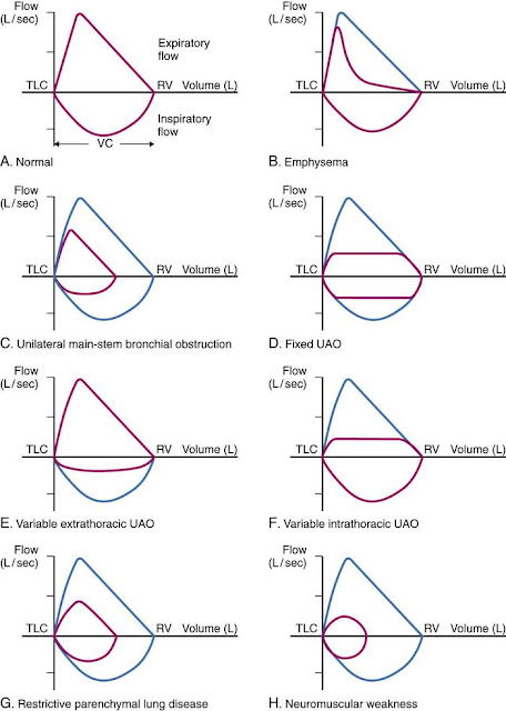
Here is a review of some of the classic flow- volume loop morphologies for pulmonary function tests.


And, lastly, here's a link to the ATS/ERS Task Force: Standardization of Lung Function Testing guide for Interpretative strategies for Lung Function Tests published in Eur Respir J in 2005.

Wednesday, August 1, 2012

Posterior Reversible Encephalopathy Syndrome

Today in Morning Report, we talked about Posterior Reversible Encephalopathy Syndrome (PRES), which is also known under the moniker, Reversible Posterior Leukoencephalopathy Syndrome (RPLS).


PRES is a neurologic syndrome diagnosed by clinical AND radiologic findings:

Clinical Presentation
Classically characterized by the following:
- headaches
- altered level of consciousness
- visual changes
- seizures

Radiologic Findings
The classic findings upon neuroimaging of PRES is symmetrical white matter edema in the posterior cerebral hemispheres, particularly the parieto-occipital regions, although this not always the case. PRES can often been seen on CT imaging of the head, although MRI brain is best.

Here's a CT head non-contrast revealing bilateral hypodensities involving the occipo-parietal region.


Here's a  T2-weighted MRI brain demonstrating multiple cortico-subcortical areas hyperintense lesions involving the occipital and parietal lobes, as well as the pons.

   
Causes of PRES
The most common causes of PRES include:
- hypertensive encephalopathy
- eclampsia
- immunosupprseive medications, including calcineurin inhibitors (cyclosporine, tacrolimus)
- TTP/HUS
- vasculitis (e.g. SLE, PAN)
- renal failure (acute and/or chronic)

Management of PRES
- The most important aspect of management is to treat the underlying cause (whether this means blood pressure lowering, delivery of baby, discontinuation of offending medication, plasmapheresis, etc.)! 
- Seizures can be treated with antiepileptic agents, most commonly phenytoin. 

Most patients demonstrate the "R" of PRES, meaning their symptoms are reversed by treating the underlying cause, often with two weeks of management, although a small minority do not make a full neurologic recovery.